Services
Ultrasound
- A non-invasive technique that evaluated various bodily structures using high-frequency sound waves.
DIAGNOSTIC TESTS

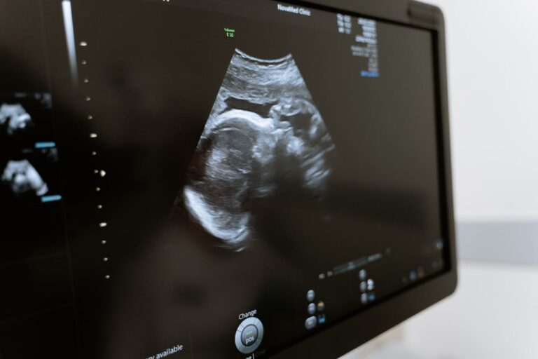
Diagnostic ultrasonography is a type of imaging that creates pictures of your body’s internal structures through the application of high-frequency sound waves. For the diagnosis and treatment of a wide range of illnesses and ailments, the pictures can offer important information.
While certain ultrasound exams require inserting a device into your body, the majority of ultrasound examinations are performed using an ultrasound device outside of your body.
The structure and movement of the body’s internal organs, as well as blood moving via blood vessels, may all be seen in real-time ultrasound imaging.
Typically, ultrasound imaging is a painless medical test that aids in the diagnosis and treatment of medical disorders by doctors.
The images from a conventional ultrasound are shown in thin, flat regions of the body. Three-dimensional (3-D) ultrasound is an advancement in ultrasound technology that converts sound wave data into three-dimensional images.
There are several uses for ultrasound, such as:
- During pregnancy, observe the uterus and ovaries and keep an eye on the health of the growing fetus.
- Identify gallbladder illness
- Analyze blood flow
- Point a needle for a biopsy or to treat a tumor.
- Look into a bump on the breast.
- Examine your thyroid gland.
- Identify prostatic and genital issues
- Examine inflammation of the joints (synovitis)
- Assess the condition of metabolic bone disease.
PRIOR TO THE UTTRASOUND
There are various kinds of ultrasonography examinations. There are tests that don’t need any preparation, tests that demand you to consume a lot of liquids before the test, and tests that demand you to fast for hours before the test. For any special instructions related to your specific test, speak with your doctor.
You might be requested to take off any jewelry from the area being inspected before your ultrasound starts. You might also be required to take off your clothes and put on a robe.
DURING THE TEST
You’ll be required to lie down on a table for inspection. Over the area of your skin that is being inspected, gel is placed. It aids in avoiding air pockets, which may obstruct the sound waves responsible for the visuals. It is simple to remove this water-based gel from skin and, if necessary, clothes.
A tiny, hand-held instrument called a transducer is pressed against the region being analyzed and moved as necessary by a skilled professional known as a sonographer to take pictures. Sound waves are sent into your body by the transducer, which then gathers and delivers the ones that bounce back to a computer to produce the photos.
Sometimes, ultrasounds are done inside your body. In this case, the transducer is attached to a probe that’s inserted into a natural opening in your body. Examples include:
Transesophageal Echocardiogram: A transducer, inserted into your esophagus, obtains heart images. It’s usually done while you are sedated.
Transrectal Ultrasound: This test creates images of the prostate by placing a special transducer into the rectum
Transvaginal Ultrasound: A special transducer is gently inserted into the vagina to get a quick look at the uterus and ovaries.
In most cases, ultrasound is painless. However, if the sonographer has to have a full bladder, you can feel some slight discomfort when the transducer is inserted into your body or guided across your body.
An ultrasound examination typically lasts thirty to sixty minutes.
FOLLOWING THE TEST
After the exam is over, you can resume your regular activities.
