Services
X-Ray Services
- A non-invasive imaging technique that evaluates various bodily structures using controlled radiation to produce detailed images of bones, tissues, and organs.
X-Ray Testing
Diagnostic X-ray imaging is a type of medical imaging that creates detailed pictures of your body’s internal structures using controlled amounts of radiation. These images provide essential information for the diagnosis and treatment of a wide range of illnesses and conditions.
Unlike some imaging techniques that require invasive procedures, X-ray imaging is entirely non-invasive and performed externally. A specialized machine directs a small amount of radiation through the body, capturing images of bones, tissues, and organs.
X-ray imaging allows healthcare providers to visualize the structure and alignment of bones, detect abnormalities in tissues, and identify issues such as fractures, infections, or tumors. The process is quick, efficient, and provides immediate results for further evaluation.
Typically, X-ray imaging is a painless and safe medical test that helps doctors diagnose and monitor medical conditions effectively. The images produced are two-dimensional, offering clear views of the body’s internal structures. Advanced X-ray technology, such as digital X-rays, enhances image quality while minimizing radiation exposure, ensuring patient safety and accurate diagnoses.
There are several uses for ultrasound, such as:
- X-rays are widely used to identify broken bones, dislocations, and other skeletal injuries.
- X-rays help detect arthritis, osteoporosis, spinal misalignment, and degenerative bone diseases.
- X-rays can reveal infections in bones (osteomyelitis) or lungs (pneumonia).
- X-rays are used to find foreign objects, such as swallowed items or embedded materials in soft tissues.
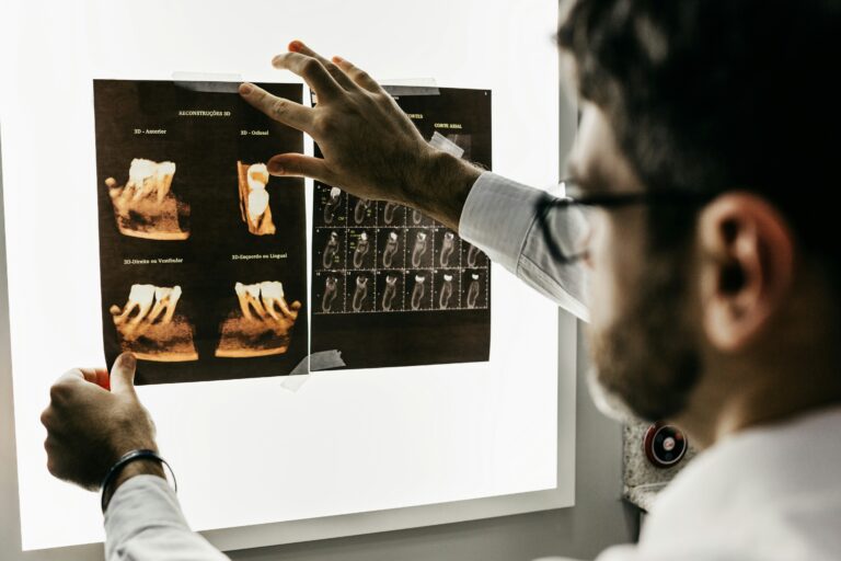
- Dentists use X-rays to examine teeth, gums, and jawbones for cavities, impacted teeth, or jaw disorders.
- X-rays help diagnose lung infections, tumors, collapsed lungs, and other chest abnormalities.
- Chest X-rays can reveal heart enlargement, fluid buildup, or vascular calcifications.
- X-rays are used during surgeries to guide the placement of implants, catheters, or other medical devices.
- X-rays are used to detect tumors in bones, lungs, and other organs, aiding in early cancer diagnosis.
PRIOR TO THE X Ray
X-ray exams vary depending on the area of the body being examined. Some tests require no special preparation, while others may need you to remove metal objects, wear loose-fitting clothing, or avoid eating or drinking beforehand. For specific instructions related to your X-ray exam, consult your doctor.
Before the procedure begins, you may be asked to remove any jewelry, eyeglasses, or metal accessories from the area being examined. You might also need to change into a medical gown.
DURING THE TEST
You will be positioned on an examination table or asked to stand, depending on the type of X-ray being performed. A trained radiologic technologist will carefully position your body to capture the required images. You may be asked to hold still and briefly hold your breath to avoid blurring the images.
A specialized X-ray machine will send a controlled amount of radiation through your body. Different tissues absorb radiation at varying levels, creating detailed images of bones and internal structures. In some cases, a contrast dye may be used to enhance visibility of certain areas, such as the digestive tract or blood vessels.
Most X-ray exams are quick and painless. However, if positioning for the X-ray requires holding an uncomfortable posture, you may experience mild discomfort.
FOLLOWING THE TEST
Once the X-ray is complete, you can typically resume your normal activities. The images will be analyzed by a radiologist, and your doctor will discuss the results with you.